


Product Information
- Product Type
- Monoclonal antibody
- Clone Number
- 3G8
- UniProt No.
- Q9NVS9
- NCBI Accession No.
- NP_006212
- Alternative names
- Protein (peptidyl-prolyl cis/trans isomerase) NIMA-interacting 1, PIN1, Peptidyl-prolyl cis-transisomerase NIMA-interacting 1, EC 5.2.1.8, Rotamase Pin1, PPIase Pin1, DOD, uBL5, PIN1,PPIase, EC 5.2.1.8, Rotamase Pin1, PPIase Pin1, Peptidyl-prolyl cis-trans isomeraseNIMA-interacting 1
Product Specification
- Host
- Mouse
- Reacts With
- Human
- Concentration
- 1mg/ml
- Formulation
- Liquid. In Phosphate-Buffered Saline (pH 7.4) with 0.02% Sodium Azide, 10% Glycerol
- Immunogen
- Recombinant human Pin1 (1-163aa) purified from E. coli
- Isotype
- IgG1 kappa
- Purification
- By protein-A affinity chromatography
- Applications
- WB, ICC/IF, IHC, FACS
- Usage
- The antibody has been tested by ELISA, Western blot, ICC/IF, FACS and IHC analysis to assure specificity and reactivity. Since application varies, however, each investigation should be titrated by the reagent to obtain optimal results.
- Storage
- Can be stored at +2C to +8C for 1 week. For long term storage, aliquot and store at -20C to -80C. Avoid repeated freezing and thawing cycles.
Data
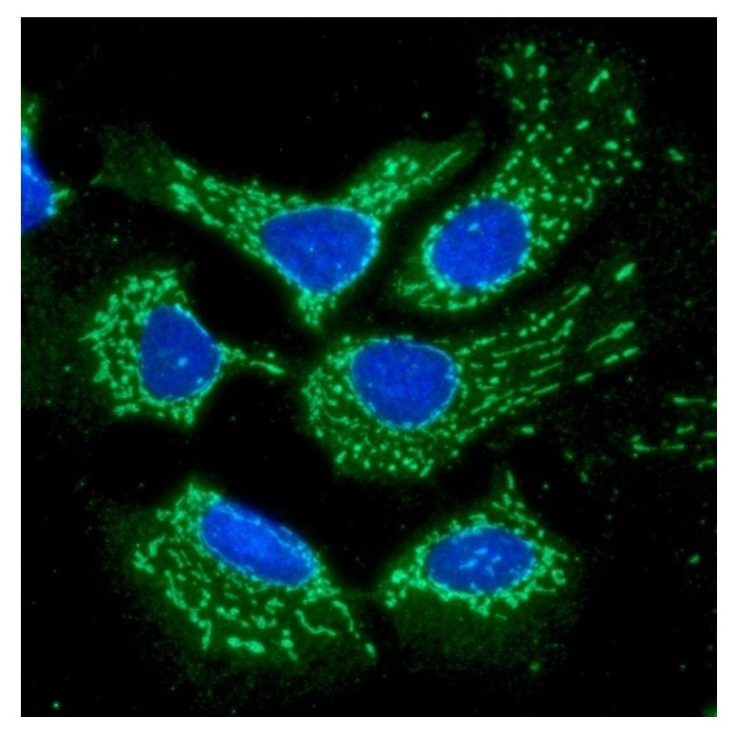
Immunflorescense (ICC/IF)

MagnifyICC/IF analysis of NM23-H1 in HeLa cells. The cell was stained with ATGA0585 (1:100). The secondary antibody (green) was used Alexa Fluor 488. DAPI was stained the cell nucleus (blue).
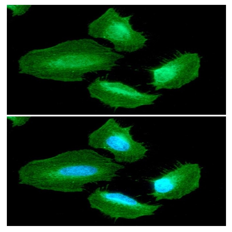
ICC/IF analysis of NM23-H1 in A549 cells. The cell was stained with ATGA0585 (1:100). The secondary antibody (green) was used Alexa Fluor 488. DAPI was stained the cell nucleus (blue).
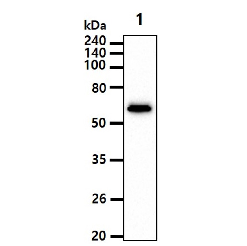
Western blot analysis (WB)

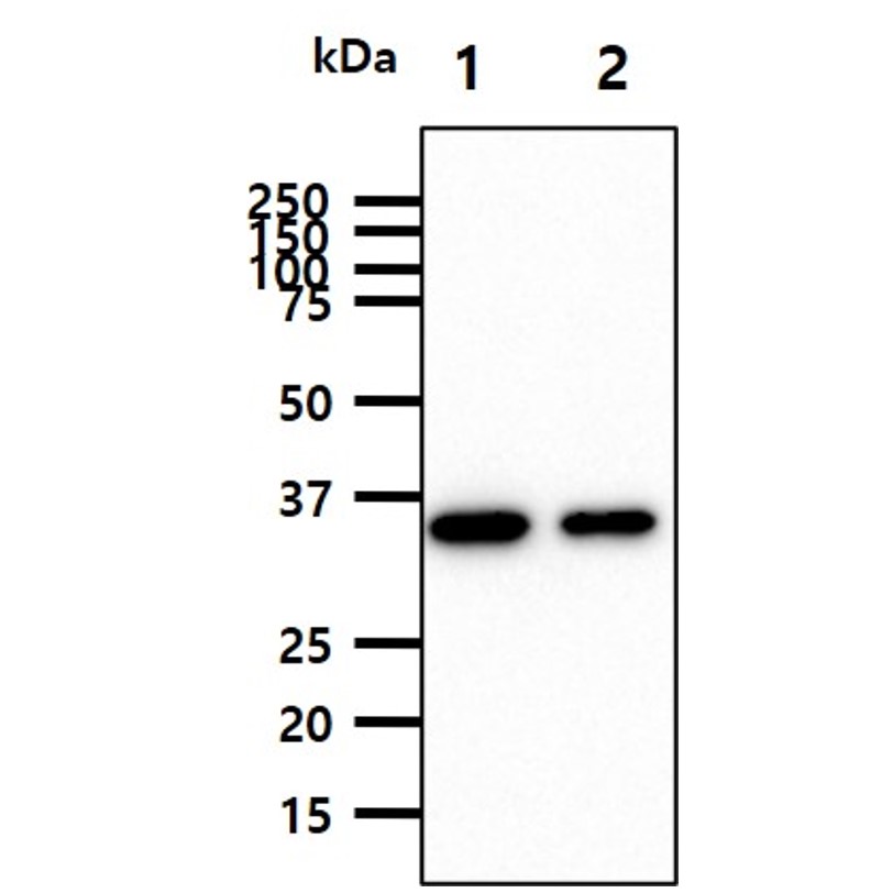
The cell lysate(40ug) was resolved by SDS-PAGE, transferred to PVDF membrane and probed with anti-human Coactosin-like Protein 1/COTL1 antibody (1:500). Proteins were visualized using a goat antimouse secondary antibody conjugated to HRP and an ECL detection system. Lane 1.: HeLa cell lysate
The Cell lysates (40ug) were resolved by SDS-PAGE, transferred to PVDF membrane and probed with anti-human PNPO antibody (1:1000). Proteins were visualized using a goat anti-mouse secondary antibody conjugated to HRP and an ECL detection system. Lane 1.: A549 cell lysate Lane 2.: HepG2 cell lysate
The recombinant protein (50ng) were resolved by SDS-PAGE, transferred to PVDF membrane and probed with anti-human NM23-H1 antibody (1:1000). Proteins were visualized using a goat anti-mouse secondary antibody conjugated to HRP and an ECL detection system. Lane 1.: Recombinant human NME1 protein Lane 2.: Recombinant human NME2 protein Lane 3.: Recombinant human NME3 protein Lane 4.: Recombinant human NME4 protein
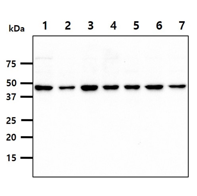
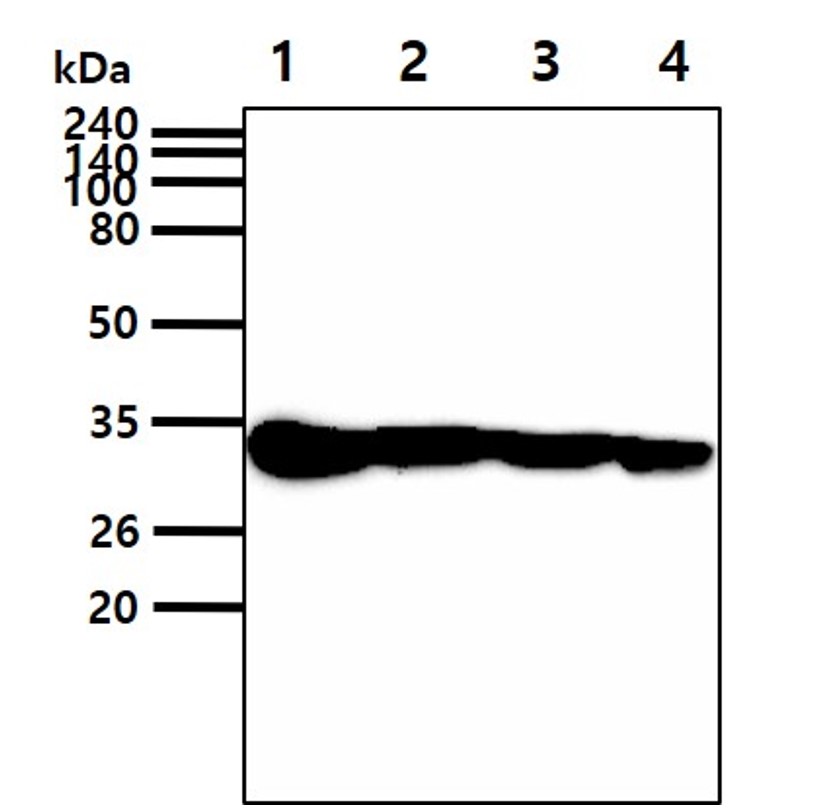
The cell lysates (40ug) were resolved by SDS-PAGE, transferred to PVDF membrane and probed with anti-human NM23-H1 antibody (1:1000). Proteins were visualized using a goat anti-mouse secondary antibody conjugated to HRP and an ECL detection system. Lane 1.: HeLa cell lysate Lane 2.: A549 cell lysate Lane 3.: Jurkat cell lysate Lane 4.: HepG2 cell lysate Lane 5.: MCF7 cell lysate Lane 6.: PC3 cell lysate
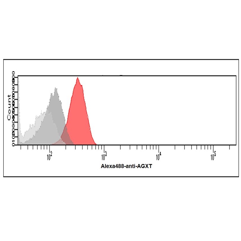
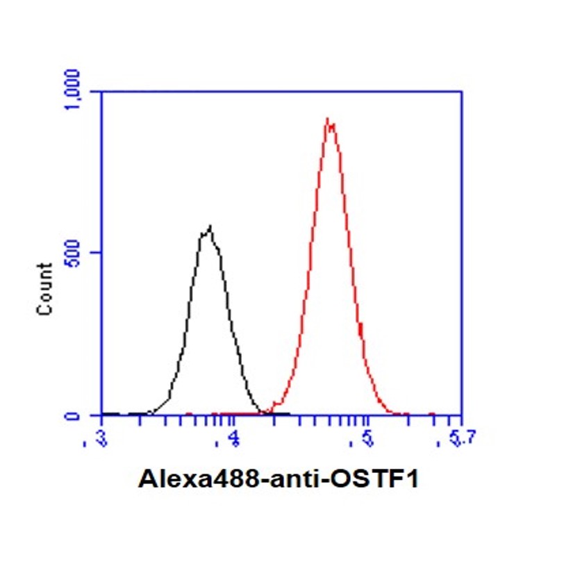
Flow cytometry (FACS)

Flow cytometry analysis of IRF5 in THP-1 cells. The cell was stained with ATGA0518 at 2-5ug for 1x10^6cells (red). A Goat anti mouse IgG (Alexa fluor 488) was used as the secondary antibody. Mouse monoclonal IgG was used as the isotype control (dark gray), cells without incubation with primary and secondary antibody was used as the negative control (light gray).

Flow cytometry analysis of Coactosin-like Protein 1/COTL1 in HeLa cells. The cell was stained with ATGA0576 at 2-5ug for 1x10^6cells (red). A Goat anti mouse IgG (Alexa fluor 488) was used as the secondary antibody. Mouse monoclonal IgG was used as the isotype control (dark gray), cells without incubation with primary and secondary antibody was used as the negative control (light gray).
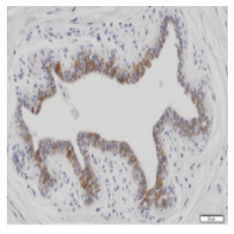
Immunohistochemistry (IHC)

Paraffin embedded sections of human cervical cancer tissue were incubated with anti-human KRT5 (1:200) for 2 hours at room temperature. Antigen retrieval was performed in 0.1M sodium citrate buffer and detected using Diaminobenzidine (DAB)
Note: For research use only. This product is not intended or approved for human, diagnostics or veterinary use.