Product Information
- Product Type
- Monoclonal Antibody
- Clone Number
- s4E5
- UniProt No.
- Q8WTS6
- NCBI Accession No.
- NP_085151
- Alternative names
- SETD7, SET7, SET9, SET7/9, SET7/9 Histone methyltransferase, SET domain-containing protein 8, SET domain-containing protein 7 FLJ21193, SET domain-containing protein 7, Lysine N-methyltransferase 7, Lysine methyltransferase, KMT7, KIAA1717, Histone-lysine N-methyltransferase SETD7, Histone-lysine N-methyltransferase, Histone lysine N methyltransferase H3 lysine 4 specific SET7, Histone lysine methyltransferase, Histone H4-K4 methyltransferase, Histone H3-K4 methyltransferase SETD7, Histone H3 lysine 4 specific methyltransferase, Histone H3 K4 methyltransferase, H4 lysine-4 specific, H3-K4-HMTase SETD7, H3 K4 HMTase, EC 2.1.1.43
Product Specification
- Host
- Mouse
- Reacts With
- Human
- Concentration
- 1mg/ml (determined by BCA assay)
- Formulation
- Liquid in. Phosphate-Buffered Saline (pH 7.4) with 0.02% Sodium Azide, 10% glycerol
- Immunogen
- Recombinant human SET 7/9 (1-366aa) purified from E. coli
- Isotype
- IgG2b kappa
- Purification
- By protein-A affinity chromatography
- Applications
- ELISA, WB, ICC/IF, FACS
- Usage
- The antibody has been tested by ELISA, Western blot, ICC/IF and FACS analysis to assure specificity and reactivity. Since application varies, however, each investigation should be titrated by the reagent to obtainoptimal results.
- Storage
- Can be stored at +2C to +8C for 1 week. For long term storage, aliquot and store at -20C to -80C. Avoid repeated freezing and thawing cycles.
Data
Immunocytochemistry/Immunofluorescence (ICC/IF)
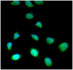
ICC/IF analysis of SET 7/9 in HeLa cells. The cell was stained with AHR0401 (1:100). The secondary antibody (green) was used Alexa Fluor 488. DAPI was stained the cell nucleus (blue).
Western blot analysis (WB)
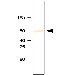
The recombinant human SET 7/9 protein was resolved by SDS-PAGE, transferred to PVDF membrane and probed with anti-human SET 7/9 antibody (1:1000). Proteins were visualized using a goat anti-mouse secondary antibody conjugated to HRP and an ECL detection system.
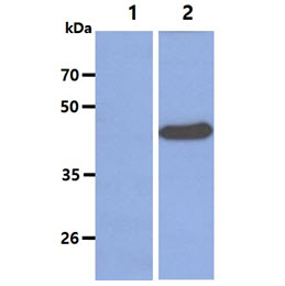
The cell lysates (5ug) were resolved by SDS-PAGE, transferred to PVDF membrane and probed with anti-human SET 7/9 antibody (1:2000). Proteins were visualized using a goat anti-mouse secondary antibody conjugated to HRP and an ECL detection system.
Lane 1.: 293T cell lysate
Lane 2.: SET 7/9 transfected 293T cell lysate
Lane 1.: 293T cell lysate
Lane 2.: SET 7/9 transfected 293T cell lysate
Flow cytometry (FACS)
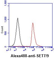
Flow cytometry analysis of SET 7/9 in Jurkat cell line, staining at 2-5ug for 1x10^6cells (red line). The secondary antibody used goat anti-mouse IgG Alexa fluor 488 conjugate. Isotype control antibody was mouse IgG (black line).
Related Publications
-
Kurash JK, et al. Methylation of p53 by Set7/9 mediates p53 acetylation and activity in vivo. (Mol Cell. 2008)
Tao Y, et al. The histone methyltransferase Set7/9 promotes myoblast differentiation and myofibril assembly. (J Cell Biol. 2011)
Han T, et al. Set7 facilitates hepatitis C virus replication via enzymatic activity-dependent attenuation of the IFN-related pathway. (J Immunol. 2015)
Zhang Y, et al. The transcription factor GATA1 and the histone methyltransferase SET7 interact to promote VEGF-mediated angiogenesis and tumor growth and predict clinical outcome of breast cancer. (Oncotarget. 2016)
Yuan H, et al. Epigenetic Histone Modifications Involved in Profibrotic Gene Regulation by 12/15-Lipoxygenase and Its Oxidized Lipid Products in Diabetic Nephropathy. (Antioxid Redox Signal. 2016)
Xu X, et al. A signature motif in LIM proteins mediates binding to checkpoint proteins and increases tumour radiosensitivity. (Nat Commun. 2017)
Note: For research use only. This product is not intended or approved for human, diagnostics or veterinary use.