Product Information
- Product Type
- Monoclonal Antibody
- Clone Number
- J1D2
- UniProt No.
- Q13158
- NCBI Accession No.
- NP_003815
- Alternative names
- Fas-associated via death domain, GIG3, MORT1, MGC8528, Fas-associated via death domain, FADD, Fas-associated via death domain FADD protein, Fas TNFRSF6 associated via death domain, Fas (TNFRSF6) associated via death domain, Fas associated via death domain, Fas associating protein, Fas associating death domain containing protein, Fas associating protein with death domain GIG 3, Growth inhibiting gene 3 protein, H sapiens mRNA for mediator of receptor induced toxicity, Mediator of receptor induced toxicity, MORT 1
Product Specification
- Host
- Mouse
- Reacts With
- Human
- Concentration
- 1mg/ml (determined by BCA assay)
- Formulation
- Liquid in. Phosphate-Buffered Saline (pH 7.4) with 0.02% Sodium Azide, 10% glycerol
- Immunogen
- Recombinant human FADD (1-208aa) purified from E. coli
- Isotype
- IgG2b kappa
- Purification
- By protein-G affinity chromatography
- Applications
- ELISA,WB,ICC/IF,IHC
- Usage
- The antibody has been tested by ELISA, Western blot, ICC/IF and IHC analysis to assure specificity and reactivity. Since application varies, however, each investigation should be titrated by the reagent to obtain optimal results.
- Storage
- Can be stored at +2C to +8C for 1 week. For long term storage, aliquot and store at -20C to -80C. Avoid repeated freezing and thawing cycles.
Data
Immunocytochemistry/Immunofluorescence (ICC/IF)
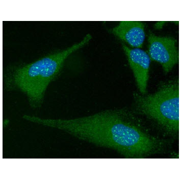
ICC/IF analysis of FADD in HeLa cells. The cell was stained with AFA0901 (1:100). The secondary antibody (green) was used Alexa Fluor 488. DAPI was stained the cell nucleus (blue).
Western blot analysis (WB)
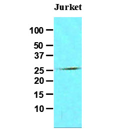
The cell lysates of Jurkat (30ug) was resolved by SDS-PAGE, transferred to NC membrane and probed with anti-human FADD (1:500). Proteins were visualized using a goat anti-mouse secondary antibody conjugated to HRP and an ECL detection system.
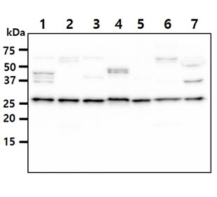
The Cell lysates (40ug) were resolved by SDS-PAGE, transferred to PVDF membrane and probed with anti-human FADD antibody (1:500). Proteins were visualized using a goat anti-mouse secondary antibody conjugated to HRP and an ECL detection system.
Lane 1. : HeLa cell lysate
Lane 2. : Raw264.7 cell lysate
Lane 3. : MCF7 cell lysate
Lane 4. : A431 cell lysate
Lane 5. : Ramos cell lysate
Lane 6. : Raji cell lysate
Lane 7. : Balb/3T3 cell lysate
Lane 1. : HeLa cell lysate
Lane 2. : Raw264.7 cell lysate
Lane 3. : MCF7 cell lysate
Lane 4. : A431 cell lysate
Lane 5. : Ramos cell lysate
Lane 6. : Raji cell lysate
Lane 7. : Balb/3T3 cell lysate
IHC Comment And Pic
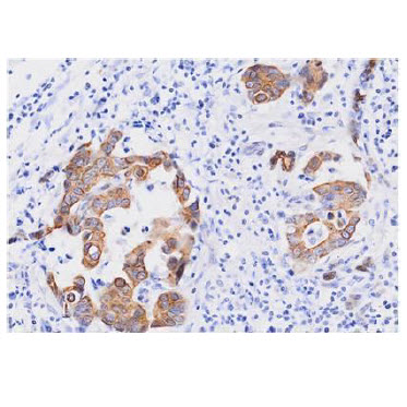
Paraffin embedded sections of human breast cancer tissue were incubated with anti-human FADD (1:50) for 2 hours at room temperature. Antigen retrieval was performed in 0.1M sodium citrate buffer and detected using Diaminobenzidine (DAB)
Note: For research use only. This product is not intended or approved for human, diagnostics or veterinary use.