

Product Information
- Product Type
- Monoclonal antibody
- Clone Number
- AT2G9
- UniProt No.
- P01111
- NCBI Accession No.
- NP_002515
- Alternative names
- Neuroblastoma RAS viral (v-ras) oncogene homolog, GTPase NRas, HRAS1, ALPS4, N-ras, NRAS1, NS6,Neuroblastoma RAS viral (v-ras) oncogene homolog OTTMuSP00000023521, AV095280, N ras, N rasprotein part 4, Neuroblastoma RAS viral (v ras) oncogene homolog, OTTHuMP00000013879,Transforming protein N Ras, v ras neuroblastoma RAS viral oncogene homolog
- Additional Information
- This product was produced from tissue culture supernatant. Ras Antibody detects endogenous levels of total K-Ras, H-Ras, and N-Ras(cell signaling).
Product Specification
- Host
- Mouse
- Reacts With
- Human
- Concentration
- 1mg/ml (determined by BCA assay)
- Formulation
- Liquid in. Phosphate-Buffered Saline (pH 7.4) with 0.02% Sodium Azide, 10% glycerol
- Immunogen
- Recombinant human NRAS (1-186aa) purified from E. coli
- Isotype
- IgG2a kappa
- Purification
- By protein-A affinity chromatography
- Applications
- ELISA, WB, FACS
- Usage
- The antibody has been tested by ELISA, Western blot and FACS analysis to assure specificity and reactivity. Since application varies, however, each investigation should be titrated by the reagent to obtain optimal results.
- Storage
- Can be stored at +2C to +8C for 1 week. For long term storage, aliquot and store at -20C to -80C. Avoid repeated freezing and thawing cycles.
Data
Western blot analysis (WB)
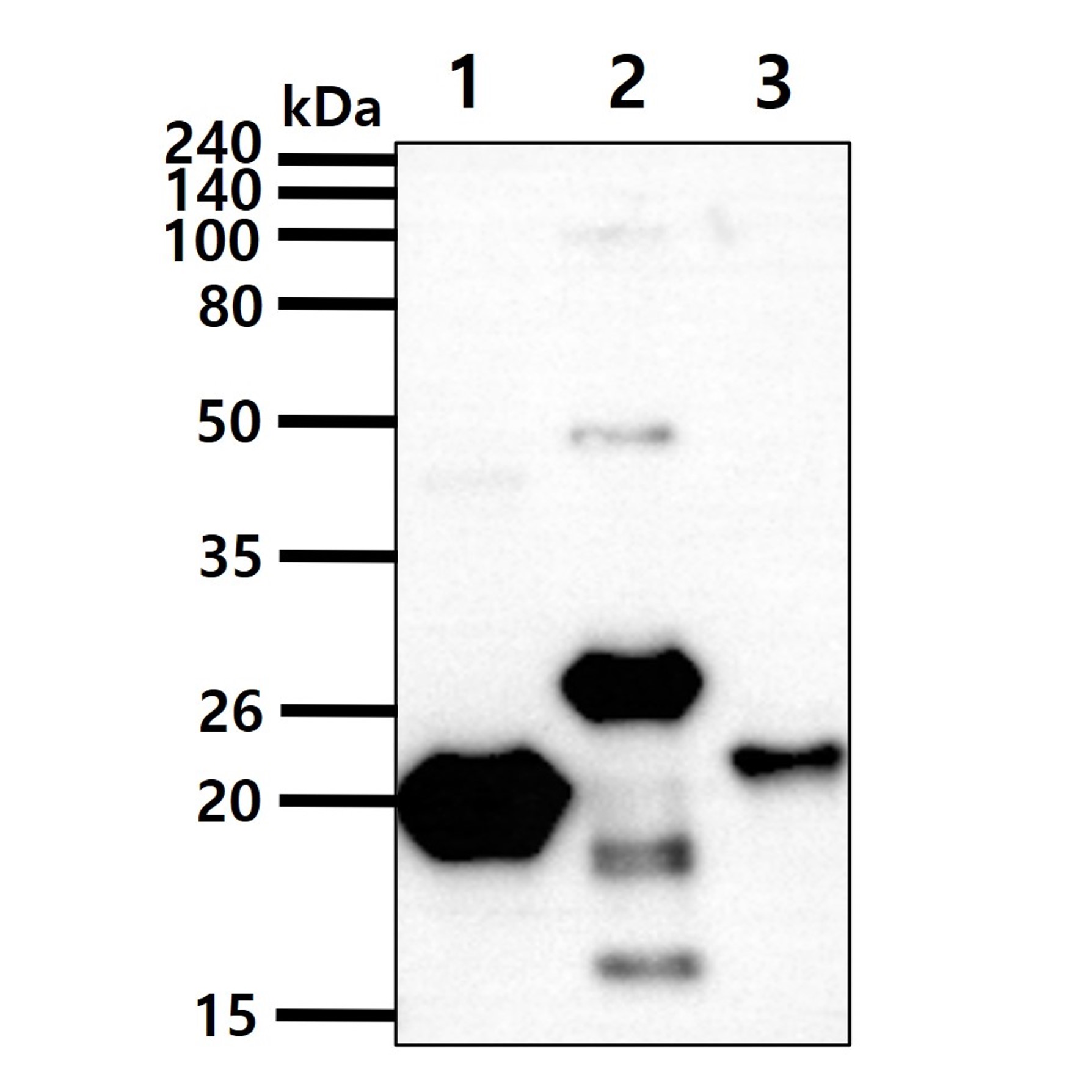
The recombinant proteins (50ng) were resolved by SDS-PAGE, transferred to PVDF membrane and probed with anti-human RAS antibody (1:1000). Proteins were visualized using a goat anti-mouse secondary antibody conjugated to HRP and an ECL detection system.
Lane 1.: Recombinant human NRAS protein
Lane 2.: Recombinant human KRAS protein
Lane 3.: Recombinant human HRAS protein
Lane 1.: Recombinant human NRAS protein
Lane 2.: Recombinant human KRAS protein
Lane 3.: Recombinant human HRAS protein
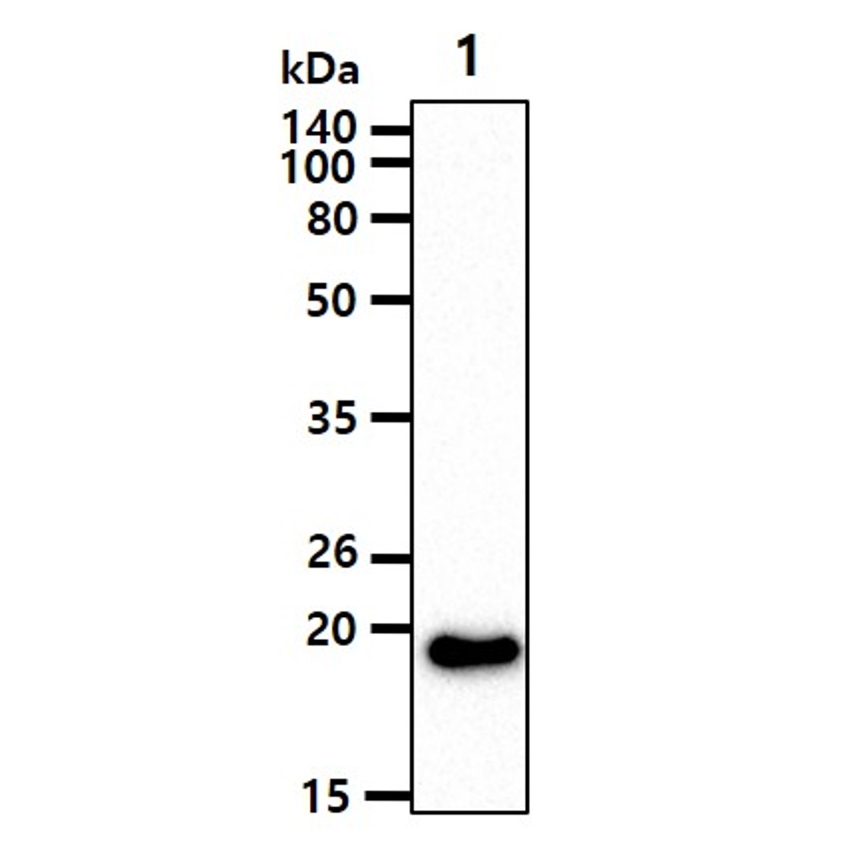
The cell lysate(40ug) was resolved by SDS-PAGE, transferred to PVDF membrane and probed with anti-human RAS antibody (1:1000). Proteins were visualized using a goat anti-mouse secondary antibody conjugated to HRP and an ECL detection system.
Lane 1.: 293T cell lysate
Lane 1.: 293T cell lysate
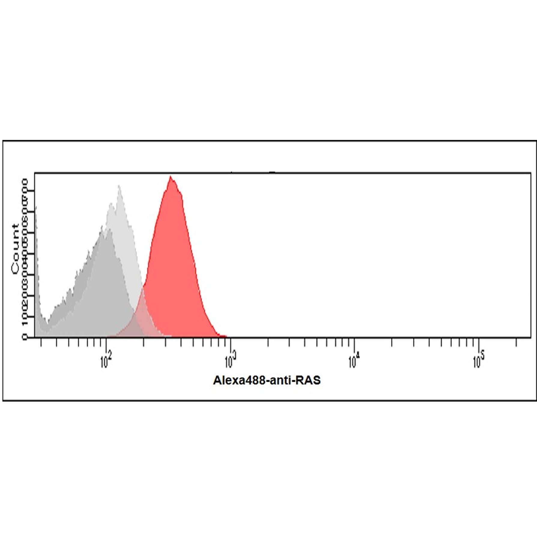
Flow cytometry (FACS)

Flow cytometry analysis of RAS in HeLa cells. The cell was stained with ATGA0573 at 2-5ug for 1x10^6cells (red). A Goat anti mouse IgG (Alexa fluor 488) was used as the secondary antibody. Mousemonoclonal IgG was used as the isotype control (dark gray), cells without incubation with primary and secondary antibody was used as the negative control (light gray).
Note: For research use only. This product is not intended or approved for human, diagnostics or veterinary use.