Product Information
- Product Type
- Monoclonal Antibody
- Clone Number
- AT38E2
- UniProt No.
- Q16611
- NCBI Accession No.
- NP_001179
- Alternative names
- BCL2-antagonist/killer 1, Apoptosis regulator BAK, BAK, BAK-LIKE, Bcl-2 homologous antagonist/killer, Bcl2-L-7, BAK-1, BAK 1, BAK like, Bak NT, BAK1, Bcl 2 homologous antagonist/killer, Bcl 2 like 7 protein, Bcl2 homologous antagonist killer, Bcl2 like 7 Protein, BCL2-antagonist/killer 1, BCL2L7, CDN 1, CDN1, Cell death inhibitor 1, MGC117255, MGC3887, NBak, Pro apoptotic protein BAK
- Additional Information
- This product was produced from tissue culture supernatant.
Product Specification
- Host
- Mouse
- Reacts With
- Human
- Concentration
- 1mg/ml (determined by BCA assay)
- Formulation
- Liquid in. Phosphate-Buffered Saline (pH 7.4) with 0.02% Sodium Azide, 10% glycerol
- Immunogen
- Recombinant human BAK (29-187aa) purified from E.coli
- Isotype
- IgG2a kappa
- Purification
- Applications
- ELISA, WB, ICC/IF, FACS
- Usage
- The antibody has been tested by ELISA, Western blot, ICC/IF and FACS to assure specificity and reactivity. Since application varies, however, each investigation should be titrated by the reagent to obtain optimal results.
- Storage
- Can be stored at +2C to +8C for 1 week. For long term storage, aliquot and store at -20C to -80C. Avoid repeated freezing and thawing cycles.
Data
Immunocytochemistry/Immunofluorescence (ICC/IF)
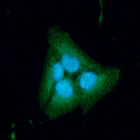
ICC/IF analysis of BAK in HeLa cells. The cell was stained with ATGA0523 (1:100). The secondary antibody (green) was used Alexa Fluor 488. DAPI was stained the cell nucleus (blue).
Western blot analysis (WB)
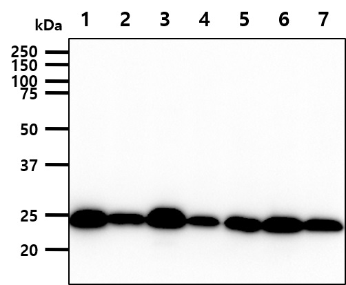
The cell lysate (40ug) were resolved by SDS-PAGE, transferred to PVDF membrane and probed with anti-human BAK antibody (1:1000). Proteins were visualized using a goat anti-mouse secondary antibody conjugated to HRP and an ECL detection system.
Lane 1 : 293T cell lysate
Lane 2 : HeLa cell lysate
Lane 3 : A431 cell lysate
Lane 4 : A549 cell lysate
Lane 5 : Jurkat cell lysate
Lane 6 : MCF7 cell lysate
Lane 7 : PC3 cell lysate
Lane 1 : 293T cell lysate
Lane 2 : HeLa cell lysate
Lane 3 : A431 cell lysate
Lane 4 : A549 cell lysate
Lane 5 : Jurkat cell lysate
Lane 6 : MCF7 cell lysate
Lane 7 : PC3 cell lysate
Flow cytometry (FACS)
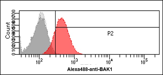
Flow cytometry analysis of BAK in HeLa cells. The cell was stained with ATGA0523 at 2-5ug for 1x10^6cells (red). A Goat anti mouse IgG (Alexa fluor 488) was used as the secondary antibody. Mouse monoclonal IgG was used as the isotype control (dark gray), cells without incubation with primary and secondary antibody was used as the negative control (light gray).
Related Publications
-
Singh A, et al. Podophyllotoxin and Rutin Modulates Ionizing Radiation-Induced Oxidative Stress and Apoptotic Cell Death in Mice Bone Marrow and Spleen. (Front Immunol. 2017)
Mebratu YA, et al. Bik reduces hyperplastic cells by increasing Bak and activating DAPk1 to juxtapose ER and mitochondria. (Nat Commun. 2017)
Liu P, et al. Downregulation of microRNA-125a is involved in intervertebral disc degeneration by targeting pro-apoptotic Bcl-2 antagonist killer 1. (Iran J Basic Med Sci. 2017)
Loo LSW, et al. BCL-xL/BCL2L1 is a critical anti-apoptotic protein that promotes the survival of differentiating pancreatic cells from human pluripotent stem cells. (Cell Death Dis. 2020)
Shen J, et al. TLR9 regulates NLRP3 inflammasome activation via the NF-kB signaling pathway in diabetic nephropathy. (Diabetol Metab Syndr. 2022)
Note: For research use only. This product is not intended or approved for human, diagnostics or veterinary use.