
Product Information
- Product Type
- Monoclonal Antibody
- Clone Number
- k2E2
- UniProt No.
- P23284
- NCBI Accession No.
- NP_000933
- Alternative names
- Peptidyl-prolyl cis-trans isomerase B, Peptidylprolyl isomerase B, PPIase B, Cphn-2, Cyclophilin B, CYP-20b, CYPB, CYP-S1, OI9, Rotamase B, S-cyclophilin, SCYLP
Product Specification
- Host
- Mouse
- Reacts With
- Human
- Concentration
- 1mg/ml (determined by BCA assay)
- Formulation
- Liquid in. Phosphate-Buffered Saline (pH 7.4) with 0.02% Sodium Azide, 10% glycerol
- Immunogen
- Recombinant human Cyclophilin B (26-216aa) purified from E. coli
- Isotype
- IgG1 kappa
- Purification
- By protein-G affinity chromatography
- Applications
- ELISA, WB, ICC/IF, FACS
- Usage
- The antibody has been tested by ELISA, Western blot, ICC/IF and FACS analysis to assure specificity and reactivity. Since application varies, however, each investigation should be titrated by the reagent to obtain optimal results.
- Storage
- Can be stored at +2C to +8C for 1 week. For long term storage, aliquot and store at -20C to -80C. Avoid repeated freezing and thawing cycles.
Data
Immunocytochemistry/Immunofluorescence (ICC/IF)
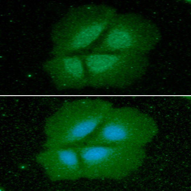
ICC/IF analysis of Cyclophilin B in Hep3B cells. The cell was stained with ACB0825 (1:100). The secondary antibody (green) was used Alexa Fluor 488. DAPI was stained the cell nucleus (blue).
Western blot analysis (WB)
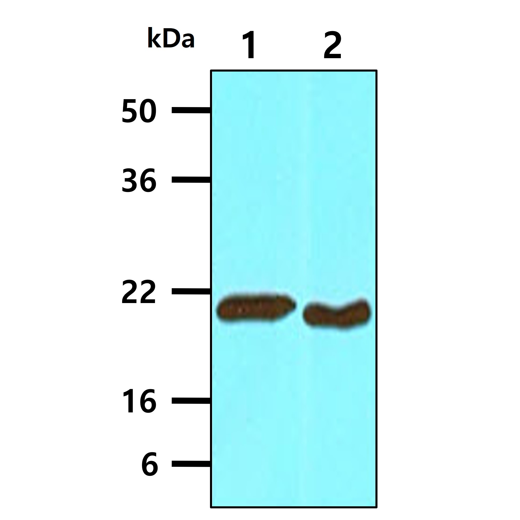
The cell lysates of HepG2 (30ug) and HeLa (30ug) were resolved by SDS-PAGE, transferred to NC membrane and probed with anti-human Cyclophilin B (1:1000). Proteins were visualized using a goat anti-mouse secondary antibody conjugated to HRP and an ECL detection system.
Lane 1.: HepG2
Lane 2.: HeLa
Lane 1.: HepG2
Lane 2.: HeLa
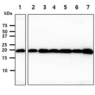
The cell lysates (40ug) were resolved by SDS-PAGE, transferred to PVDF membrane and probed with anti-human Cyclophilin B antibody (1:1000). Proteins were visualized using a goat anti-mouse secondary antibody conjugated to HRP and an ECL detection system.
Lane 1.: Jurkat cell lysate
Lane 2.: K562 cell lysate
Lane 3.: 293T cell lysate
Lane 4.: A549 cell lysate
Lane 5.: MCF7 cell lysate
Lane 6.: SK-OV-3 cell lysate
Lane 7.: LnCap cell lysate
Lane 1.: Jurkat cell lysate
Lane 2.: K562 cell lysate
Lane 3.: 293T cell lysate
Lane 4.: A549 cell lysate
Lane 5.: MCF7 cell lysate
Lane 6.: SK-OV-3 cell lysate
Lane 7.: LnCap cell lysate
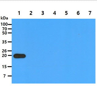
The recombinant proteins (50ng) were resolved by SDS-PAGE, transferred to PVDF membrane and probed with anti-human Cyclophilin B antibody (1:1000). Proteins were visualized using a goat anti-mouse secondary antibody conjugated to HRP and an ECL detection system.
Lane 1.: Cyclophilin B recombinant protein
Lane 2.: Cyclophilin A recombinant protein
Lane 3.: Cyclophilin D recombinant protein
Lane 4.: Cyclophilin E recombinant protein
Lane 5.: Cyclophilin F recombinant protein
Lane 6.: Cyclophilin G recombinant protein
Lane 7.: Cyclophilin H recombinant protein
Flow cytometry (FACS)
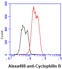
Flow cytometry analysis of Cyclophilin B in Hep3B cell line, staining at 2-5ug for 1x10^6cells (red line). The secondary antibody used goat anti-mouse IgG Alexa fluor 488 conjugate. Isotype control antibody was mouse IgG (black line).
Related Publications
-
Choi JW, et al. Severe osteogenesis imperfecta in cyclophilin B-deficient mice. (PLoS Genet. 2009)
Dubnikov T, et al. PrP-containing aggresomes are cytosolic components of an ER quality control mechanism. (J Cell Sci. 2016)
Mukadam AS, et al. Analysis of novel endosome-to-Golgi retrieval genes reveals a role for PLD3 in regulating endosomal protein sorting and amyloid precursor protein processing. (Cell Mol Life Sci. 2018)
Note: For research use only. This product is not intended or approved for human, diagnostics or veterinary use.