Product Information
- Product Type
- Monoclonal Antibody
- Clone Number
- p6c7
- UniProt No.
- P35813
- NCBI Accession No.
- NP_066283
- Alternative names
- Pyruvate dehydrogenase phosphatase catalytic subunit 1, Pyruvate dehydrogenase acetyl-transferring-phosphatase 1, Protein phosphatase Mg2+/Mn2+dependent 1A, Protein phosphatase 2C, Protein phosphatase 1A (formerly 2C) magnesium-dependent alpha isoform, Protein phosphatase 1A (formerly 2C), PPM2C, PPM1A, PP2CA, PP2C alpha, PDPC 1, PDP 1, PDP, MGC9201, FLJ42306, EC 3.1.3.43
Product Specification
- Host
- Mouse
- Reacts With
- Human
- Concentration
- 1mg/ml (determined by BCA assay)
- Formulation
- Liquid in. Phosphate-Buffered Saline (pH 7.4) with 0.02% Sodium Azide, 10% glycerol
- Immunogen
- Recombinant human PP2Calpha (1-382aa) purified from E. coli
- Isotype
- IgG2b kappa
- Purification
- By protein-G affinity chromatography
- Applications
- ELISA, WB, ICC/IF
- Usage
- The antibody has been tested by ELISA, Western blot and ICC/IF analysis to assure specificity and reactivity. Since application varies, however, each investigation should be titrated by the reagent to obtainoptimal results.
- Storage
- Can be stored at +2C to +8C for 1 week. For long term storage, aliquot and store at -20C to -80C. Avoid repeated freezing and thawing cycles.
Data
Immunocytochemistry/Immunofluorescence (ICC/IF)
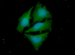
ICC/IF analysis of PP2C alpha in HeLa cells. The cell was stained with APP0401 (1:100). The secondary antibody (green) was used Alexa Fluor 488. DAPI was stained the cell nucleus (blue).
Western blot analysis (WB)
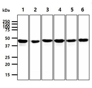
The cell lysates (40ug) were resolved by SDS-PAGE, transferred to PVDF membrane and probed with anti-human PP2C alpha antibody (1:1000). Proteins were visualized using a goat anti-mouse secondary antibody conjugated to HRP and an ECL detection system.
Lane 1.: Jurkat cell lysate
Lane 2.: HeLa cell lysate
Lane 3.: K562 cell lysate
Lane 4.: MCF7 cell lysate
Lane 5.: A549 cell lysate
Lane 6.: Raji cell lysate
Lane 1.: Jurkat cell lysate
Lane 2.: HeLa cell lysate
Lane 3.: K562 cell lysate
Lane 4.: MCF7 cell lysate
Lane 5.: A549 cell lysate
Lane 6.: Raji cell lysate
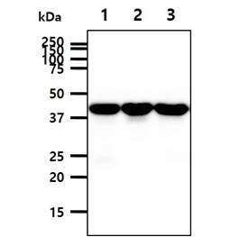
The tissue lysates (40ug) were resolved by SDS-PAGE, transferred to PVDF membrane and probed with anti-human PP2C alpha antibody (1:1000). Proteins were visualized using a goat anti-mouse secondary antibody conjugated to HRP and an ECL detection system.
Lane 1.: Mouse kidney tissue lysate
Lane 2.: Mouse brain tissue lysate
Lane 3.: Mouse liver tissue lysate
Lane 1.: Mouse kidney tissue lysate
Lane 2.: Mouse brain tissue lysate
Lane 3.: Mouse liver tissue lysate
Related Publications
-
Kim JS, et al. The interaction of hepatitis B virus X protein and protein phosphatase type 2 Calpha and its effect on IL-6. (Biochem Biophys Res Commun. 2006)
Kim YG, et al. Role of protein phosphatase magnesium-dependent 1A and anti-protein phosphatase magnesium-dependent 1A autoantibodies in ankylosing spondylitis. (Arthritis Rheumatol. 2014)
Pereira JM, et al. Infection Reveals a Modification of SIRT2 Critical for Chromatin Association. (Cell Rep. 2018)
Note: For research use only. This product is not intended or approved for human, diagnostics or veterinary use.