Product Information
- Product Type
- Monoclonal Antibody
- Clone Number
- 2A5
- UniProt No.
- P04792
- NCBI Accession No.
- NP_001531
- Alternative names
- Heat shock 27 kDa protein, Hspb1, Hsp25, Hsp27, Hsp27, HspB1, Heat shock 27 kDa protein, HSP 27, Growth-related 25 kDa protein, P25, HSP25, Heat shock protein beta-1, Heat shock 27 kDa protein 28 kDa heat shock protein, CMT2F, DKFZp586P1322, Estrogen regulated 24 kDa protein, at shock 25kDa protein 1, Heat shock 27kDa protein 1, Heat shock 28kDa protein 1, Heat Shock Protein 27, Heat shock protein beta 1, Heat Shock Protein27, HS.76067, Hsp 28, Hsp B1, Hsp28, HspB1, SRP27, Stress responsive protein 27.
Product Specification
- Host
- Mouse
- Reacts With
- Human
- Concentration
- 1mg/ml (determined by BCA assay)
- Formulation
- Liquid in. Phosphate-Buffered Saline (pH 7.4) with 0.02% Sodium Azide, 10% glycerol
- Immunogen
- Recombinant human Hsp27 (1-205aa) purified from E. coli
- Isotype
- IgG1 kappa
- Purification
- By protein-G affinity chromatography
- Applications
- ELISA, WB, ICC/IF, IHC, FACS
- Usage
- The antibody has been tested by ELISA, Western blot, ICC/IF, FACS and IHC analysis to assure specificity and reactivity. Since application varies, however, each investigation should be titrated by the reagent to obtain optimal results.
- Storage
- Can be stored at +2C to +8C for 1 week. For long term storage, aliquot and store at -20C to -80C. Avoid repeated freezing and thawing cycles.
Data
Immunocytochemistry/Immunofluorescence (ICC/IF)
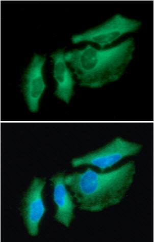
ICC/IF analysis of Hsp27 in HeLa cells. The cell was stained with AHS0702 (1:100). The secondary antibody (green) was used Alexa Fluor 488. DAPI was stained the cell nucleus (blue).
Western blot analysis (WB)
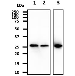
The cell lysates (40ug) were resolved by SDS-PAGE, transferred to PVDF membrane and probed with anti-human Hsp27 (1:1,000). Proteins were visualized using a goat anti-mouse secondary antibody conjugated to HRP and an ECL detection system.
Lane 1.: HeLa cell lysate
Lane 2.: K562 cell lysate
Lane 3.: MCF7 cell lysate
Lane 1.: HeLa cell lysate
Lane 2.: K562 cell lysate
Lane 3.: MCF7 cell lysate
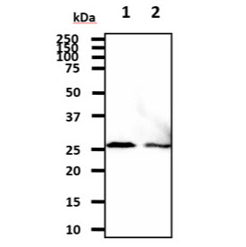
The cell lysates (40ug) were resolved by SDS-PAGE, transferred to PVDF membrane and probed with anti-human Hsp27 (1:1,000). Proteins were visualized using a goat anti-mouse secondary antibody conjugated to HRP and an ECL detection system.
Lane 1.: A431 cell lysate
Lane 2.: HepG2 cell lysate
Lane 1.: A431 cell lysate
Lane 2.: HepG2 cell lysate
Flow cytometry (FACS)
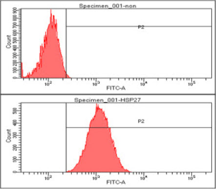
Flow cytometry analysis of Hsp27 in A549 cell line, staining at 2-5ug for 1x10^6cells. The secondary antibody used goat anti-mouse IgG Alexa fluor 488 conjugate.
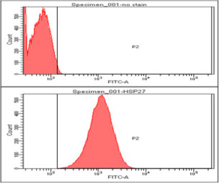
Flow cytometry analysis of Hsp27 in HeLa cell line, staining at 2-5ug for 1x10^6cells. The secondary antibody used goat anti-mouse IgG Alexa fluor 488 conjugate.
Immunohistochemistry (IHC)
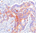
Paraffin embedded sections of human colon cancer tissue were incubated with anti-human Hsp27 (1:50) for 2 hours at room temperature. Antigen retrieval was performed in 0.1M sodium citrate buffer and detected using Diaminobenzidine (DAB)
Related Publications
-
Kaiser F, et al. Association between circulating levels of heat-shock protein 27 and aggressive periodontitis. (Cell Stress Chaperones. 2018)
Eickholz P, et al. Effect of nonsurgical periodontal therapy on haematological parameters in grades B and C periodontitis: an exploratory analysis. (Clin Oral Investig. 2020)
Note: For research use only. This product is not intended or approved for human, diagnostics or veterinary use.