


Product Information
- Product Type
- Monoclonal Antibody
- Clone Number
- AT18E6
- UniProt No.
- Q96AD5
- NCBI Accession No.
- NP_065109
- Alternative names
- Adipose triglyceride lipase, Calcium-independent phospholipase A2, Desnutrin IPLA2-zeta, Pigment epithelium-derived factor receptor, TTS2.2, Transport-secretion protein 2, TTS2, ATGL, Patatin-like phospholipase domain-containing protein 2, PNPLA2
- Additional Information
- ATGA0135 has been replaced with a catalog number ATGA0122.
Product Specification
- Host
- Mouse
- Reacts With
- Human
- Concentration
- 1mg/ml (determined by BCA assay)
- Formulation
- Liquid in. Phosphate-Buffered Saline (pH 7.4) with 0.02% Sodium Azide, 10% glycerol
- Immunogen
- Recombinant human ATGL (30-504aa) purified from E. coli
- Isotype
- IgG2b kappa
- Purification
- By protein-G affinity chromatography
- Applications
- ELISA, WB, ICC/IF, FACS
- Usage
- The antibody has been tested by ELISA, Western blot, ICC/IF and FACS analysis to assure specificity and reactivity. Since application varies, however, each investigation should be titrated by the reagent to obtain optimal results.
- Storage
- Can be stored at +2C to +8C for 1 week. For long term storage, aliquot and store at -20C to -80C. Avoid repeated freezing and thawing cycles.
Data
Immunocytochemistry/Immunofluorescence (ICC/IF)
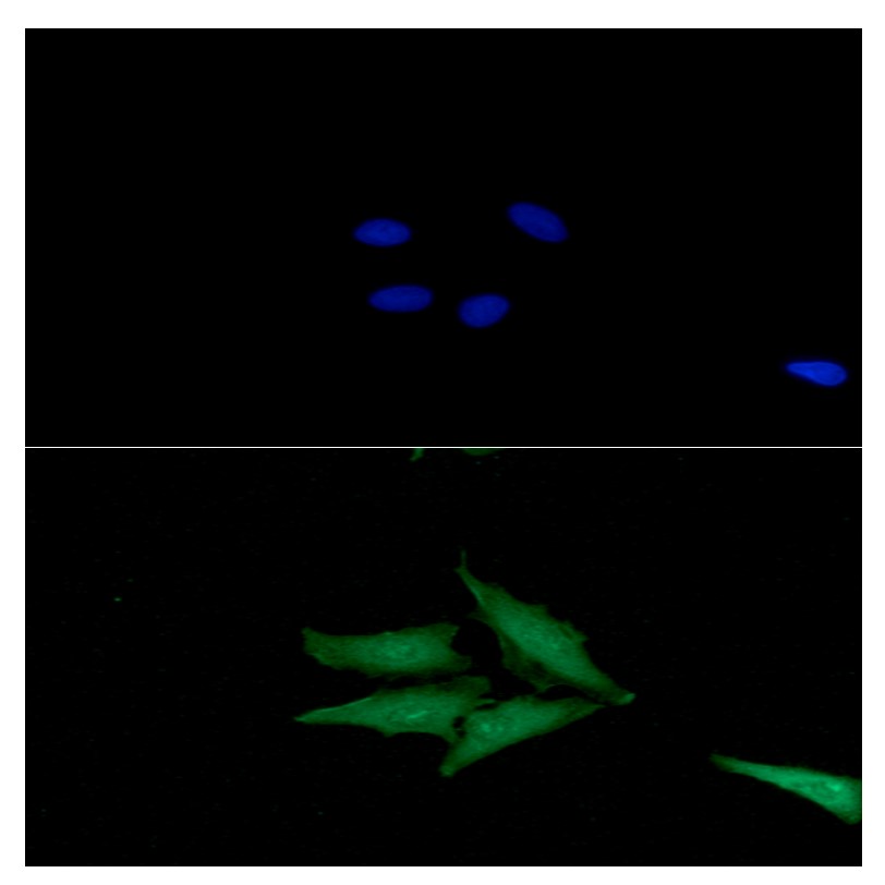
ICC/IF analysis of ATGL in Hep3B cells. The cell was stained with ATGA0122 (1:100). The secondary antibody (green) was used Alexa Fluor 488. DAPI was stained the cell nucleus (blue).

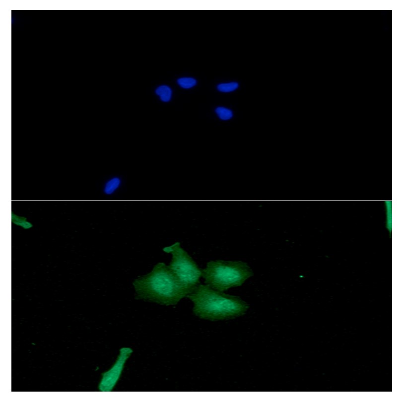
ICC/IF analysis of ATGL in HeLa cells. The cell was stained with ATGA0122 (1:100). The secondary antibody (green) was used Alexa Fluor 488. DAPI was stained the cell nucleus (blue).

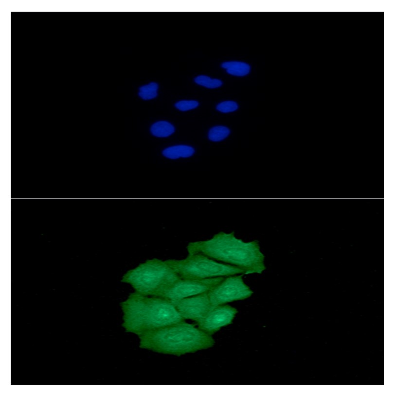
ICC/IF analysis of ATGL in A549 cells. The cell was stained with ATGA0122 (1:100). The secondary antibody (green) was used Alexa Fluor 488. DAPI was stained the cell nucleus (blue).
Western blot analysis (WB)
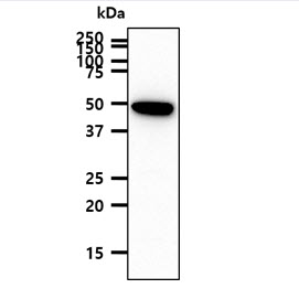
The A431 cell lysate (40ug) was resolved by SDS-PAGE, transferred to PVDF membrane and probed with anti-human ATGL antibody (1:1000). Proteins were visualized using a goat anti-mouse secondary antibody conjugated to HRP and an ECL detection system.
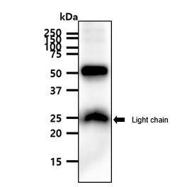
The mouse adipose tissue lysate (40ug) was resolved by SDS-PAGE, transferred to PVDF membrane and probed with anti-human ATGL antibody (1:1000). Proteins were visualized using a goat anti-mouse secondary antibody conjugated to HRP and an ECL detection system.
Flow cytometry (FACS)

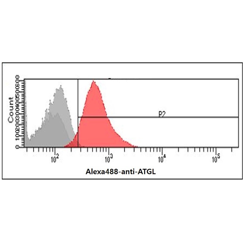
Flow cytometry analysis of ATGL in Hep3B cells. The cell was stained with ATGA0122 at 2-5ug for 1x10^6cells (red). A Goat anti mouse IgG (Alexa fluor 488) was used as the secondary antibody. Mouse monoclonal IgG was used as the isotype control (dark gray), cells without incubation with primary and secondary antibody was used as the negative control (light gray).
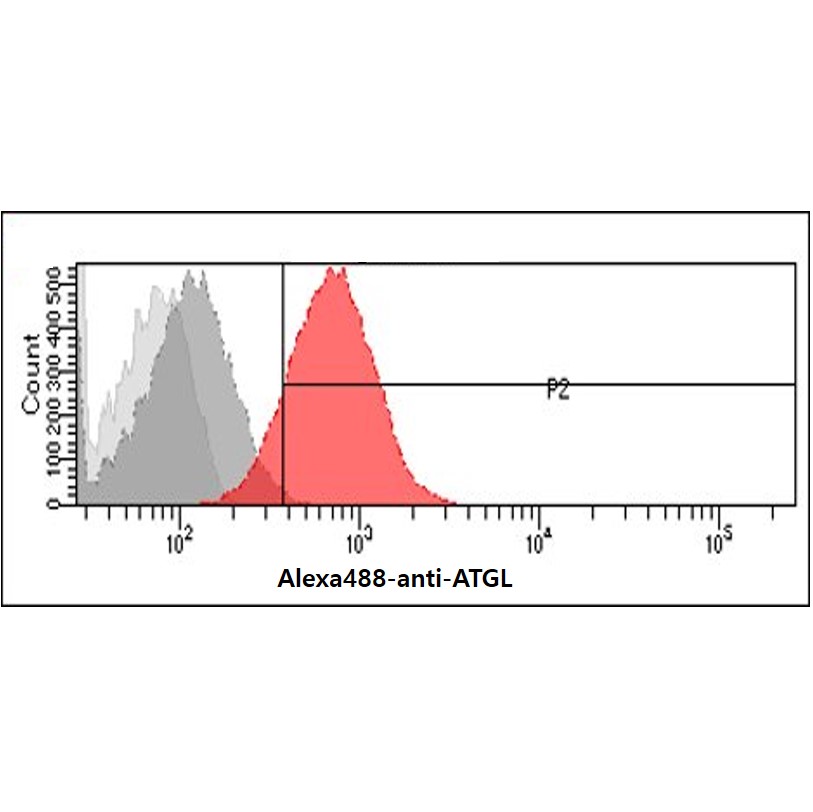
Flow cytometry analysis of ATGL in HeLa cells. The cell was stained with ATGA0122 at 2-5ug for 1x10^6cells (red). A Goat anti mouse IgG (Alexa fluor 488) was used as the secondary antibody. Mouse monoclonal IgG was used as the isotype control (dark gray), cells without incubation with primary and secondary antibody was used as the negative control (light gray).
Note: For research use only. This product is not intended or approved for human, diagnostics or veterinary use.