Product Information
- Product Type
- Monoclonal Antibody
- Clone Number
- AT3B2
- UniProt No.
- P10916
- NCBI Accession No.
- NP_000423
- Alternative names
- Slow cardiac myosin regulatory light chain 2, MLC2, CMH10, DKFZp779C0562, Slow cardiac myosin regulatory light chain 2, MYL2, Slow cardiac myosin regulatory light chain 2 Cardiac myosin light chain-2, MLC 2v, MYL 2, Cardiac ventricular myosin light chain 2, RLC of myosin, Myosin light chain 2 regulatory cardiac slow, Myosin light polypeptide 2 regulatory cardiac slow, Myosin regulatory light chain 2 ventricular cardiac muscle isoform, Myosin regulatory light chain 2 ventricular/cardiac muscle isoform, Regulatory light chain of myosin
Product Specification
- Host
- Mouse
- Reacts With
- Human
- Concentration
- 1mg/ml (determined by BCA assay)
- Formulation
- Liquid in. Phosphate-Buffered Saline (pH 7.4) with 0.02% Sodium Azide, 10% glycerol
- Immunogen
- Recombinant human MYL2 (1-166aa) purified from E. coli
- Isotype
- IgG2b kappa
- Purification
- By protein-A affinity chromatography
- Applications
- ELISA, WB, ICC/IF, FACS
- Usage
- The antibody has been tested by ELISA, Western blot, ICC/IF and FACS analysis to assure specificity and reactivity. Since application varies, however, each investigation should be titrated by the reagent to obtain optimal results.
- Storage
- Can be stored at +2C to +8C for 1 week. For long term storage, aliquot and store at -20C to -80C. Avoid repeated freezing and thawing cycles.
Data
Immunocytochemistry/Immunofluorescence (ICC/IF)
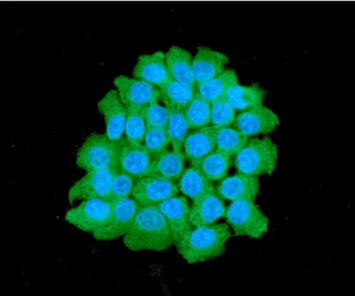
ICC/IF analysis of MYL2 in A431 cells. The cell was stained with ATGA0151 (1:100). The secondary antibody (green) was used Alexa Fluor 488. DAPI was stained the cell nucleus (blue).
Western blot analysis (WB)
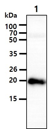
The tissue lysate (40ug) was resolved by SDS-PAGE, transferred to PVDF membrane and probed with anti-human MYL2 antibody (1:1000). Proteins were visualized using a goat anti-mouse secondary antibody conjugated to HRP and an ECL detection system.
Lane 1.: Mouse heart tissue lysate
Lane 1.: Mouse heart tissue lysate
Flow cytometry (FACS)
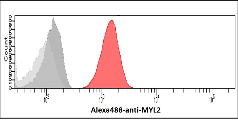
Flow cytometry analysis of MYL2 in A431 cells. The cell was stained with ATGA0151 at 2-5ug for 1x10^6cells (red). A Goat anti mouse IgG (Alexa fluor 488) was used as the secondary antibody. Mouse monoclonal IgG was used as the isotype control (dark gray), cells without incubation with primary and secondary antibody was used as the negative control (light gray).
Related Publications
-
Beveridge RD, et al. The leukemia-associated Rho guanine nucleotide exchange factor LARG is required for efficient replication stress signaling. (Cell Cycle. 2014)
Russo V, et al. Porous, Ventricular Extracellular Matrix-Derived Foams as a Platform for Cardiac Cell Culture. (Biores Open Access. 2015)
Mourino-Alvarez L, et al. A comprehensive study of calcific aortic stenosis: from rabbit to human samples. (Dis Model Mech. 2018)
Acharya BR, et al. A Mechanosensitive RhoA Pathway that Protects Epithelia against Acute Tensile Stress. (Dev Cell. 2018)
Note: For research use only. This product is not intended or approved for human, diagnostics or veterinary use.