
Product Information
- Product Type
- Monoclonal Antibody
- Clone Number
- s4E5
- UniProt No.
- Q8WTS6
- NCBI Accession No.
- NP_085151
- Alternative names
- SETD7, SET7, SET9, SET7/9, SET7/9 Histone methyltransferase, SET domain-containing protein 8, SET domain-containing protein 7 FLJ21193, SET domain-containing protein 7, Lysine N-methyltransferase 7, Lysine methyltransferase, KMT7, KIAA1717, Histone-lysine N-methyltransferase SETD7, Histone-lysine N-methyltransferase, Histone lysine N methyltransferase H3 lysine 4 specific SET7, Histone lysine methyltransferase, Histone H4-K4 methyltransferase, Histone H3-K4 methyltransferase SETD7, Histone H3 lysine 4 specific methyltransferase, Histone H3 K4 methyltransferase, H4 lysine-4 specific, H3-K4-HMTase SETD7, H3 K4 HMTase, EC 2.1.1.43
- Additional Information
- This product was produced from tissue culture supernatant.
Product Specification
- Host
- Mouse
- Reacts With
- Human
- Concentration
- 1mg/ml (determined by BCA assay)
- Formulation
- Liquid in. Phosphate-Buffered Saline (pH 7.4) with 0.02% Sodium Azide, 10% glycerol
- Immunogen
- Recombinant human SET7/9 (1-366aa) purified from E. coli
- Isotype
- IgG2b kappa
- Purification
- By protein-A affinity chromatography
- Applications
- ELISA, WB, ICC/IF, FACS
- Usage
- The antibody has been tested by ELISA, Western blot, ICC/IF and FACS analysis to assure specificity and reactivity. Since application varies, however, each investigation should be titrated by the reagent to obtain optimal results
- Storage
- Can be stored at +2C to +8C for 1 week. For long term storage, aliquot and store at -20C to -80C. Avoid repeated freezing and thawing cycles.
Data
Immunocytochemistry/Immunofluorescence (ICC/IF)
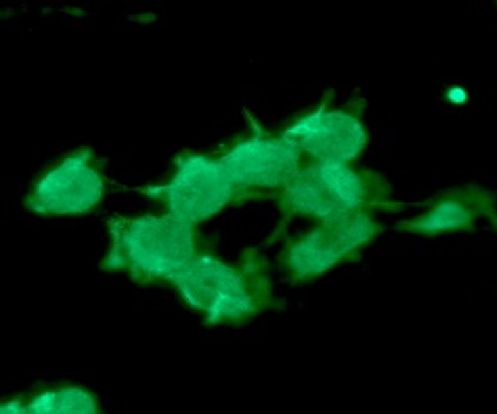
ICC/IF analysis of SET7/9 in HeLa cells. The cell was stained with ATGA0524 (1:100). The secondary antibody (green) was used Alexa Fluor 488.
Western blot analysis (WB)
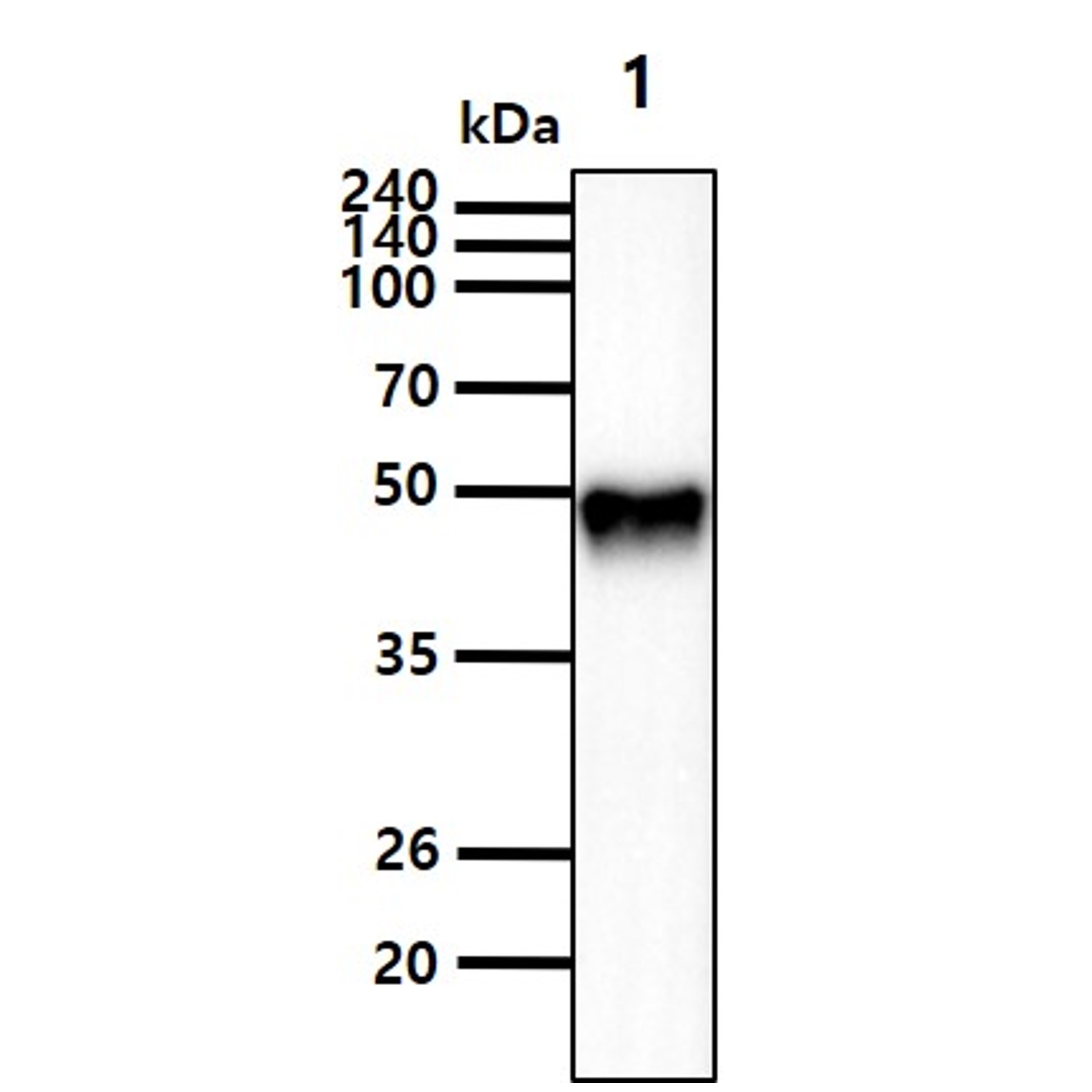
The Recombinant protein(50ng) was resolved by SDS-PAGE, transferred to PVDF membrane and probed with anti-human SET7/9 antibody (1:1000). Proteins were visualized using a goat anti-mouse secondary antibody conjugated to HRP and an ECL detection system.
Lane 1.: Recombinant human SET7/9 protein
Lane 1.: Recombinant human SET7/9 protein

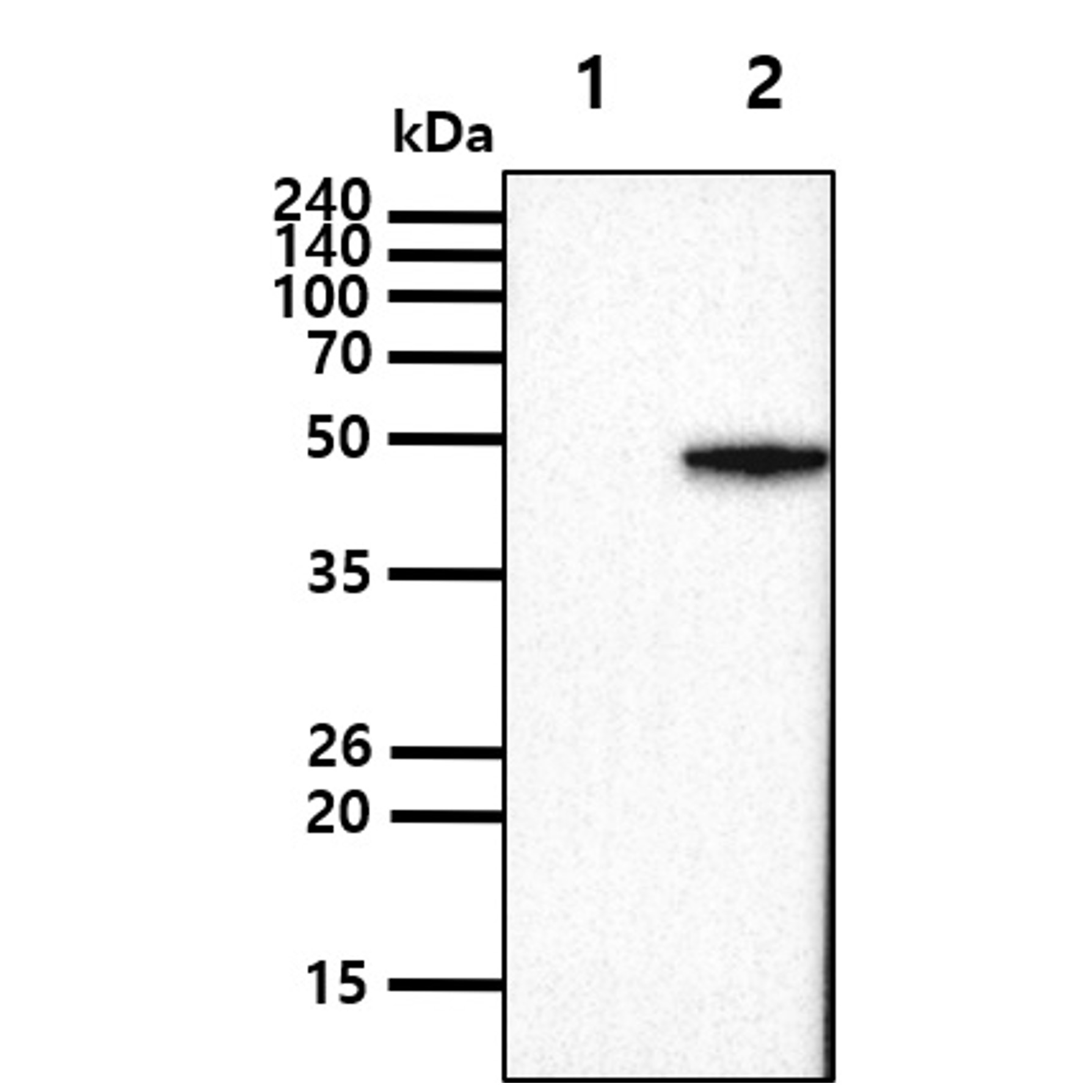
The Cell lysates (5ug) were resolved by SDS-PAGE, transferred to PVDF membrane and probed with anti-human SET7/9 antibody (1:2000). Proteins were visualized using a goat anti-mouse secondary antibody conjugated to HRP and an ECL detection system.
Lane 1.: 293T cell lysate
Lane 2.: SET7/9 transfected 293T cell lysate
Lane 1.: 293T cell lysate
Lane 2.: SET7/9 transfected 293T cell lysate
Flow cytometry (FACS)
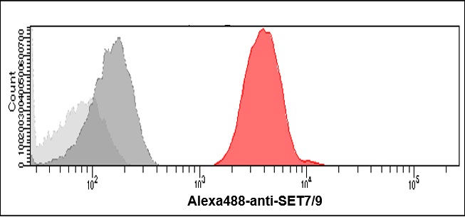
Flow cytometry analysis of SET7/9 in Jurkat cells. The cell was stained with ATGA0524 at 2-5ug for 1x10^6cells (red). A Goat anti mouse IgG (Alexa fluor 488) was used as the secondary antibody. Mouse monoclonal IgG was used as the isotype control (darkgray), cells without incubation with primary and secondary antibody was used as the negative control (light gray).
Related Publications
-
Giannios I, et al. Protein Methyltransferase Inhibition Decreases Endocrine Specification Through the Upregulation of Aldh1b1 Expression. (Stem Cells. 2019)
Si W, et al. SET7/9 promotes multiple malignant processes in breast cancer development via RUNX2 activation and is negatively regulated by TRIM21. (Cell Death Dis. 2020)
Xie H, et al. METTL3/YTHDF2 m6 A axis promotes tumorigenesis by degrading SETD7 and KLF4 mRNAs in bladder cancer. (J Cell Mol Med. 2020)
Cao L, et al. Downregulation of SETD7 promotes migration and invasion of lung cancer cells via JAK2/STAT3 pathway. (Int J Mol Med. 2020)
Jetton TL, et al. SetD7 (Set7/9) is a novel target of PPARγ that promotes the adaptive pancreatic β-cell glycemic response. (J Biol Chem. 2021)
Li X, et al. Associating Preoperative MRI Features and Gene Expression Signatures of Early-stage Hepatocellular Carcinoma Patients using Machine Learning. (J Clin Transl Hepatol. 2022)
Li J, et al. ZKSCAN5 Activates VEGFC Expression by Recruiting SETD7 to Promote the Lymphangiogenesis, Tumour Growth, and Metastasis of Breast Cancer. (Front Oncol. 2022)
Note: For research use only. This product is not intended or approved for human, diagnostics or veterinary use.