Product Information
- Product Type
- Monoclonal antibody
- Clone Number
- AT4E8
- UniProt No.
- P46406
- NCBI Accession No.
- NP_001075722.1
- Alternative names
- Glyceraldehyde-3-phosphate dehydrogenase isoform 1, Peptidyl-cysteine S-nitrosylase GAPDH, GAPD, G3PD
- Additional Information
- This product was produced from tissue culture supernatant.
Product Specification
- Host
- Mouse
- Concentration
- 1mg/ml (determined by BCA assay)
- Formulation
- Liquid in. Phosphate-Buffered Saline (pH 7.4) with 0.02% Sodium Azide, 10% glycerol
- Immunogen
- GAPDH from rabbit muscle
- Isotype
- IgG2b kappa
- Purification
- By protein-A affinity chromatography
- Applications
- ELISA, WB, ICC/IF
- Usage
- The antibody has been tested by ELISA, Western blot and ICC/IF analysis to assure specificity and reactivity. Since application varies, however, each investigation should be titrated by the reagent to obtain optimal results.
- Storage
- Can be stored at +2C to +8C for 1 week. For long term storage, aliquot and store at -20C to -80C. Avoid repeated freezing and thawing cycles.
Data
Immunocytochemistry/Immunofluorescence (ICC/IF)
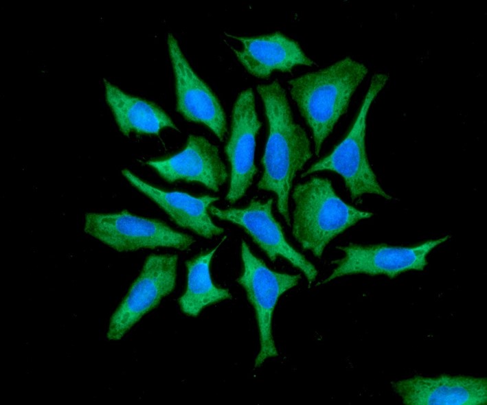
ICC/IF analysis of GAPDH in HeLa cells. The cell was stained with ATGA0559 (1:100). The secondary antibody (green) was used Alexa Fluor 488. DAPI was stained the cell nucleus (blue).
Western blot analysis (WB)
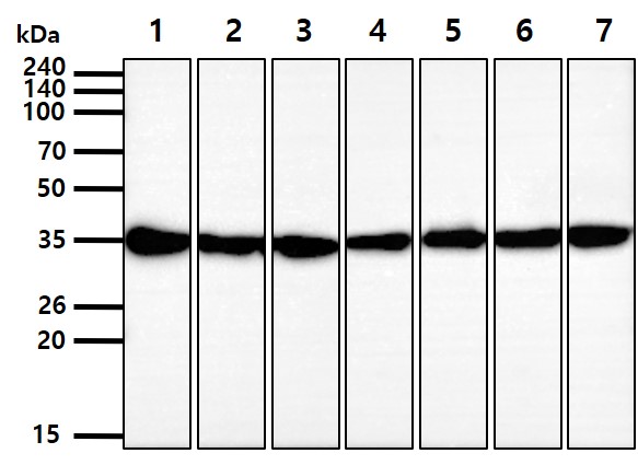
The cell lysates (40ug) were resolved by SDS-PAGE, transferred to PVDF membrane and probed with ant-GAPDH antibody (1:1000). Proteins were visualized using a goat anti-mouse secondary antibody conjugated to HRP and an ECL detection system.
Lane 1.: HepG2 cell lysate
Lane 2.: MCF7 cell lysate
Lane 3.: Jurkat cell lysate
Lane 4.: K562 cell lysate
Lane 5.: A431 cell lysate
Lane 6.: LNCap cell lysate
Lane 7.: Ramos cell lysate
Lane 1.: HepG2 cell lysate
Lane 2.: MCF7 cell lysate
Lane 3.: Jurkat cell lysate
Lane 4.: K562 cell lysate
Lane 5.: A431 cell lysate
Lane 6.: LNCap cell lysate
Lane 7.: Ramos cell lysate
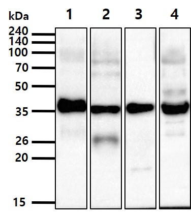
The tissue lysates (20ug) were resolved by SDS-PAGE, transferred to PVDF membrane and probedwith anti-GAPDH antibody (1:1000). Proteins were visualized using a goat anti-mouse secondary antibody conjugated to HRP and an ECL detection system.
Lane 1.: Mouse Brain tissue lysate
Lane 2.: Mouse Spleen tissue lysate
Lane 3.: Mouse Eye tissue lysate
Lane 4.: Mouse Muscle tissue lysate
Lane 1.: Mouse Brain tissue lysate
Lane 2.: Mouse Spleen tissue lysate
Lane 3.: Mouse Eye tissue lysate
Lane 4.: Mouse Muscle tissue lysate
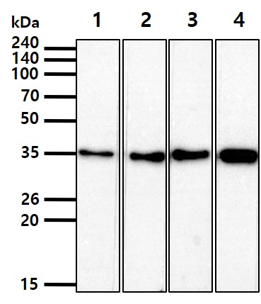
The 293T cell lysate (40ug) were resolved by SDS-PAGE, transferred to PVDF membrane and probed with anti-GAPDH. Proteins were visualized using a goat anti-mouse secondary antibody conjugated to HRPand an ECL detection system.
Lane 1.: Anti-GAPDH monoclonal antibody (1:100,000)
Lane 2.: Anti-GAPDH monoclonal antibody (1:50,000)
Lane 3.: Anti-GAPDH monoclonal antibody (1:10,000)
Lane 4.: Anti-GAPDH monoclonal antibody (1:1,000)
Lane 1.: Anti-GAPDH monoclonal antibody (1:100,000)
Lane 2.: Anti-GAPDH monoclonal antibody (1:50,000)
Lane 3.: Anti-GAPDH monoclonal antibody (1:10,000)
Lane 4.: Anti-GAPDH monoclonal antibody (1:1,000)
Related Publications
-
Chang Y, et al. Clinical impact of serum exosomal microRNA in liver fibrosis. (PLoS One. 2021)
Hah YS, et al. β-Sitosterol Attenuates Dexamethasone-Induced Muscle Atrophy via Regulating FoxO1-Dependent Signaling in C2C12 Cell and Mice Model. (Nutrients. 2022)
Hah YS, et al. Rutin Prevents Dexamethasone-Induced Muscle Loss in C2C12 Myotube and Mouse Model by Controlling FOXO3-Dependent Signaling. (Antioxidants. 2023)
Park SW, et al. Development of new tools to study membrane-anchored mammalian Atg8 proteins. (Autophagy. 2023)
Note: For research use only. This product is not intended or approved for human, diagnostics or veterinary use.