Product Information
- Product Type
- Monoclonal Antibody
- Clone Number
- AT4G6
- UniProt No.
- P60709
- NCBI Accession No.
- NP_001092
- Alternative names
- Actin, cytoplasmic 1, Beta-actin
Product Specification
- Host
- Mouse
- Reacts With
- Human
- Concentration
- 0.2mg/ml (determined by BCA assay)
- Formulation
- Liquid in. Phosphate-Buffered Saline (pH 7.4) with 0.02% Sodium Azide, 10% glycerol
- Immunogen
- Recombinant human ACTB (1-375aa) purified from E. coli
- Isotype
- IgG1 kappa
- Purification
- By protein-A affinity chromatography
- Applications
- ELISA, WB, ICC/IF
- Usage
- The antibody has been tested by ELISA, Western blot and ICC/IF analysis to assure specificity and reactivity. Since application varies, however, each investigation should be titrated by the reagent to obtain optimal results.
- Storage
- Can be stored at +2C to +8C for 1 week. For long term storage, aliquot and store at -20C to -80C. Avoid repeated freezing and thawing cycles.
Data
Immunocytochemistry/Immunofluorescence (ICC/IF)
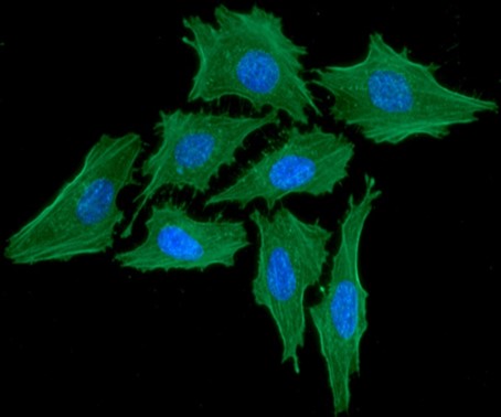
ICC/IF analysis of ACTB in HeLa cells. The cell was stained with ATGA0570 (1:100). The secondary antibody (green) was used Alexa Fluor 488. DAPI was stained the cell nucleus (blue).
Western blot analysis (WB)
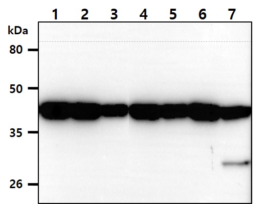
The cell lysates (40ug) were resolved by SDS-PAGE, transferred to PVDF membrane and probed with anti-human ACTB antibody (1:1000). Proteins were visualized using a goat anti-mouse secondary antibody conjugated to HRP and an ECL detection system.
Lane 1.: HeLa cell lysate
Lane 2.: Jurkat cell lysate
Lane 3.: PC3 cell lysate
Lane 4.: HepG2 cell lysate
Lane 5.: MCF7 cell lysate
Lane 6.: U87-MG cell lysate
Lane 7.: NIH-3T3 cell lysate
Lane 1.: HeLa cell lysate
Lane 2.: Jurkat cell lysate
Lane 3.: PC3 cell lysate
Lane 4.: HepG2 cell lysate
Lane 5.: MCF7 cell lysate
Lane 6.: U87-MG cell lysate
Lane 7.: NIH-3T3 cell lysate
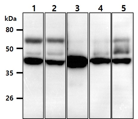
The mouse tissue lysates (40ug) were resolved by SDS-PAGE, transferred to PVDF membrane and probed with anti-human ACTB antibody (1:1000). Proteins were visualized using a goat anti-mouse secondary antibody conjugated to HRP and an ECL detection system.
Lane 1.: Kidney tissue lysate
Lane 2.: Liver tissue lysate
Lane 3.: Spleen tissue lysate
Lane 4.: Bone marrow tissue lysate
Lane 5.: Stomach tissue lysate
Lane 1.: Kidney tissue lysate
Lane 2.: Liver tissue lysate
Lane 3.: Spleen tissue lysate
Lane 4.: Bone marrow tissue lysate
Lane 5.: Stomach tissue lysate
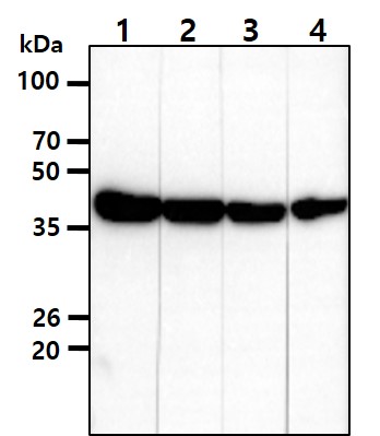
The HeLa cell lysates (40ug) were resolved by SDS-PAGE, transferred to PVDF membrane and probed with anti-human ACTB antibody. Proteins were visualized using a goat anti-mouse secondary antibody conjugated to HRP and an ECL detection system.
Lane 1.: ACTB antibody 1:1000
Lane 2.: ACTB antibody 1:3000
Lane 3.: ACTB antibody 1:5000
Lane 4.: ACTB antibody 1:10000
Lane 1.: ACTB antibody 1:1000
Lane 2.: ACTB antibody 1:3000
Lane 3.: ACTB antibody 1:5000
Lane 4.: ACTB antibody 1:10000
Related Publications
-
Yang YJ et al., Liquid Chromatography/Tandem Mass Spectrometry Analysis of Sophora flavescens Aiton and Protective Effects against Alcohol-Induced Liver Injury and Oxidative Stress in Mice. (Antioxidants (Basel). 2024)
Yang YJ et al., Korean Mistletoe (Viscum album var. coloratum) Ethanol Extracts Enhance Intestinal Barrier Function and Alleviate Inflammation. (Antioxidants (Basel) 2025)
Kim MJ et al., Potential Chondroprotective Effect of Artemisia annua L. Water Extract on SW1353 Cell. (Int. J. Mol. Sci. 2025)
Han JY et al., Cytoplasmic HMGB1 promotes the activation of JAK2-STAT3 signaling and PD-L1 expression in breast cancer. (Mol Med. 2025)
Kim MJ et al., Anti-inflammatory and antioxidant properties of Camellia sinensis L. extract as a potential therapeutic for atopic dermatitis through NF-κB pathway inhibition. (Sci Rep. 2025)
Note: For research use only. This product is not intended or approved for human, diagnostics or veterinary use.