
Product Information
- Product Type
- Monoclonal Antibody
- Clone Number
- 2E9
- UniProt No.
- P10809
- NCBI Accession No.
- NP_002147
- Alternative names
- Heat shock 60kDa protein 1, CPN60, GroEL, HSP65, SPG13, HuCHA60, Heat shock 60kDa protein 1, HSP60, Heat shock 60kDa protein 1 60 kDa chaperonin, GroEL, E, coli, homolog of, 60 kDa heat shock protein mitochondrial, 60kDa, cb863, Chaperonin, Chaperonin 60, Chaperonin, 60-KD, CPN 60, fa04a05, fb22d10, fi27b05, GroEL Homolog, Heat shock 60kD protein 1 (chaperonin), Heat shock 60kD protein 1 chaperonin, heat shock 60kDa protein 1 (chaperonin), Heat shock protein 1 (chaperonin), Heat Shock Protein 60, Heat shock protein 65, HLD4, Hsp 60, HSP 65, HSPD 1, HSPD1, HuCHA60, id:ibd2197, Spastic paraplegia 13 Mitochondrial matrix protein P1, P60 lymphocyte protein, sb:cb144, Short heat shock protein 60 Hsp60s1, Spastic paraplegia 13 (autosomal dominant), SPG 13, wu:fa04a05, wu:fb22d10, wu:fi04a12, wu:fi27b05.
Product Specification
- Host
- Mouse
- Reacts With
- Human
- Concentration
- 1mg/ml (determined by BCA assay)
- Formulation
- Liquid in. Phosphate-Buffered Saline (pH 7.4) with 0.02% Sodium Azide, 10% glycerol
- Immunogen
- Recombinant human Hsp60 (1-573aa) purified from E. coli
- Isotype
- IgG1 kappa
- Purification
- By protein-G affinity chromatography
- Applications
- ELISA, WB, ICC/IF, IHC
- Usage
- The antibody has been tested by ELISA, Western blot, ICC/IF and IHC analysis to assure specificity and reactivity. Since application varies, however, each investigation should be titrated by the reagent to obtain optimal results.
- Storage
- Can be stored at +2C to +8C for 1 week. For long term storage, aliquot and store at -20C to -80C. Avoid repeated freezing and thawing cycles.
Data
Immunocytochemistry/Immunofluorescence (ICC/IF)
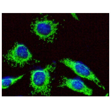
ICC/IF analysis of Hsp60 in HeLa cells. The cell was stained with AHS0815 (1:100). The secondary antibody (green) was used Alexa Fluor 488. DAPI was stained the cell nucleus (blue).
Western blot analysis (WB)
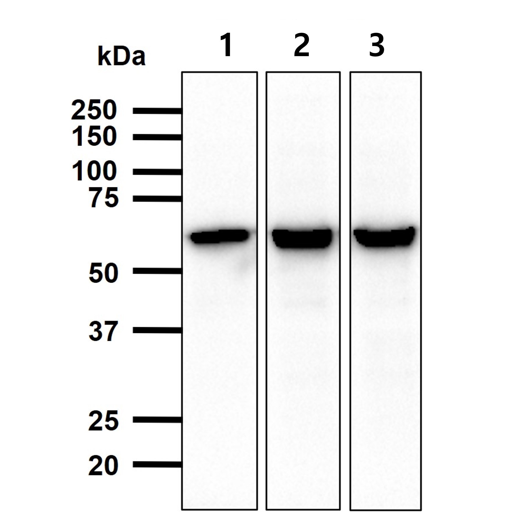
Cell lysates (40ug) were resolved by SDS-PAGE, transferred to PVDF membrane and probed with anti-human Hsp60 (1:1000). Proteins were visualized using a goat anti-mouse secondary antibody conjugated to HRP and an ECL detection system.
Lane 1.: 293T cell lysate
Lane 2.: HeLa cell lysate
Lane 3.: A549 cell lysate
Lane 1.: 293T cell lysate
Lane 2.: HeLa cell lysate
Lane 3.: A549 cell lysate
Immunohistochemistry (IHC)
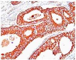
Paraffin embedded sections of human colon cancer tissue were incubated with anti-human Hsp60 (1:50) for 2 hours at room temperature. Antigen retrieval was performed in 0.1M sodium citrate buffer and detected using Diaminobenzidine (DAB)
Note: For research use only. This product is not intended or approved for human, diagnostics or veterinary use.