Product Information
- Product Type
- Monoclonal Antibody
- Clone Number
- AT2G9
- UniProt No.
- P01111
- NCBI Accession No.
- NP_002515
- Alternative names
- Neuroblastoma RAS viral (v-ras) oncogene homolog, GTPase NRas, HRAS1, ALPS4, N-ras, NRAS1, NS6, Neuroblastoma RAS viral (v-ras) oncogene homolog OTTMuSP00000023521, AV095280, N ras, N ras protein part 4, Neuroblastoma RAS viral (v ras) oncogene homolog, OTTHuMP00000013879, Transforming protein N Ras, v ras neuroblastoma RAS viral oncogene homolog
- Additional Information
- Ras Antibody detects endogenous levels of total K-Ras, H-Ras, and N-Ras(cell signaling).
Product Specification
- Host
- Mouse
- Reacts With
- Human
- Concentration
- 1mg/ml (determined by BCA assay)
- Formulation
- Liquid in. Phosphate-Buffered Saline (pH 7.4) with 0.02% Sodium Azide, 10% glycerol
- Immunogen
- Recombinant human NRAS (1-186aa) purified from E. coli
- Isotype
- IgG2a kappa
- Purification
- By protein-G affinity chromatography
- Applications
- ELISA, WB, ICC/IF, FACS
- Usage
- The antibody has been tested by ELISA, Western blot, ICC/IF and FACS analysis to assure specificity and reactivity. Since application varies, however, each investigation should be titrated by the reagent to obtain optimal results.
- Storage
- Can be stored at +2C to +8C for 1 week. For long term storage, aliquot and store at -20C to -80C. Avoid repeated freezing and thawing cycles.
Data
Immunocytochemistry/Immunofluorescence (ICC/IF)
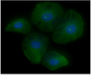
ICC/IF analysis of RAS in HeLa cells. The cell was stained with ATGA0203 (1:100). The secondary antibody (green) was used Alexa Fluor 488. DAPI was stained the cell nucleus (blue).
Western blot analysis (WB)
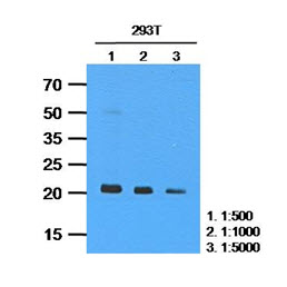
The cell lysates of 293T(35ug) were resolved by SDS-PAGE, transferred to PVDF membrane and probed with anti-human RAS (1:500~1:5000). Proteins were visualized using a goat anti-mouse secondary antibody conjugated to HRP and an ECL detection system.
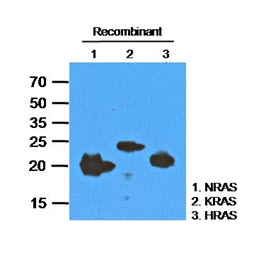
The recombinant proteins (200ng) were resolved by SDS-PAGE, transferred to PVDF membrane and probed with anti-human RAS antibody (1:1000). Proteins were visualized using a goat anti-mouse secondary antibody conjugated to HRP and an ECL detection system.
Lane 1.: Recombinant human NRAS protein
Lane 2.: Recombinant human KRAS protein
Lane 3.: Recombinant human HRAS protein
Lane 1.: Recombinant human NRAS protein
Lane 2.: Recombinant human KRAS protein
Lane 3.: Recombinant human HRAS protein
Flow cytometry (FACS)
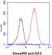
Flow cytometry analysis of RAS in HeLa cell line, staining at 2-5ug for 1x10^6cells (red line). The secondary antibody used goat anti-mouse IgG Alexa fluor 488 conjugate. Isotype control antibody was mouse IgG (black line).
Note: For research use only. This product is not intended or approved for human, diagnostics or veterinary use.