Product Information
- Product Type
- Monoclonal Antibody
- Clone Number
- AT3G11
- UniProt No.
- P17174
- NCBI Accession No.
- NP_002070
- Alternative names
- Glutamic-oxaloacetic transaminase 1, Aspartate aminotransferase 1, Aspartate transaminase 1, AST1, SGOT, AST, cAspAT, Cysteine aminotransferase cytoplasmic, Cysteine transaminase, Cytoplasmic, cCAT, Transaminase A
Product Specification
- Host
- Mouse
- Reacts With
- Human
- Concentration
- 1mg/ml (determined by BCA assay)
- Formulation
- Liquid in. Phosphate-Buffered Saline (pH 7.4) with 0.02% Sodium Azide, 10% glycerol
- Immunogen
- Recombinant human GOT1 (1-413aa) purified from E. coli
- Isotype
- IgG2a kappa
- Purification
- By protein-A affinity chromatography
- Applications
- ELISA, WB, ICC/IF, FACS
- Usage
- The antibody has been tested by ELISA, Western blot analysis, Flow cytometry and ICC/IF to assure specificity and reactivity. Since application varies, however, each investigation should be titrated by the reagent to obtain optimal results.
- Storage
- Can be stored at +2C to +8C for 1 week. For long term storage, aliquot and store at -20C to -80C. Avoid repeated freezing and thawing cycles.
Data
Immunocytochemistry/Immunofluorescence (ICC/IF)
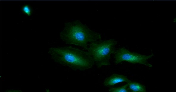
ICC/IF analysis of GOT1 in A549 cells line, stained with DAPI (Blue) for nucleus staining and monoclonal anti-human GOT1 antibody (1:100) with goat anti-mouse IgG-Alexa fluor 488 conjugate (Green).
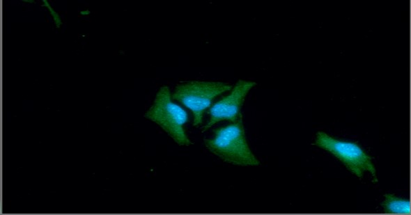
ICC/IF analysis of GOT1 in HeLa cells line, stained with DAPI (Blue) for nucleus staining and monoclonal anti-human GOT1 antibody (1:100) with goat anti-mouse IgG-Alexa fluor 488 conjugate (Green).
Western blot analysis (WB)
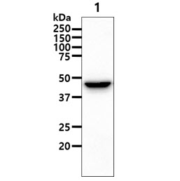
The cell lysate (40ug) were resolved by SDS-PAGE, transferred to PVDF membrane and probed with anti-human GOT1 antibody (1:1000). Proteins were visualized using a goat anti-mouse secondary antibody conjugated to HRP and an ECL detection system.
Lane 1 : A427 cell lysate
Lane 1 : A427 cell lysate
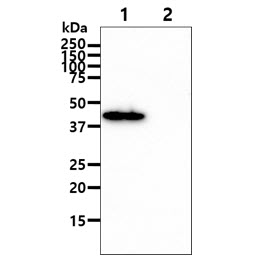
The recombinat proteins (100ng) were resolved by SDS-PAGE, transferred to PVDF membrane and probed with anti-human GOT1 antibody (1:1000). Proteins were visualized using a goat anti-mouse secondary antibody conjugated to HRP and an ECL detection system.
Lane 1 : Recombinant human GOT1
Lane 2 : Recombinant human GOT2
Lane 1 : Recombinant human GOT1
Lane 2 : Recombinant human GOT2
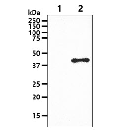
The cell lysates (10ug) were resolved by SDS-PAGE, transferred to PVDF membrane and probed with anti-human GOT1 antibody (1:1000). Proteins were visualized using a goat anti-mouse secondary antibody conjugated to HRP and an ECL detection system.
Lane 1 : 293T cell lysate
Lane 2 : GOT1 Transfected 293T cell lysate
Lane 1 : 293T cell lysate
Lane 2 : GOT1 Transfected 293T cell lysate
Flow cytometry (FACS)
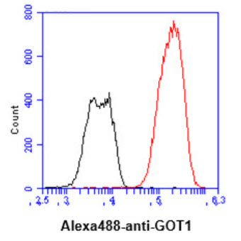
Flow cytometry analysis of GOT1 in Hep3B cell line, staining at 2-5ug for 1x10⁶cells (red line). The secondary antibody used goat anti-mouse IgG Alexa fluor 488 conjugate. Isotype control antibody was mouse IgG (black line).
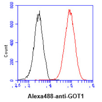
Flow cytometry analysis of GOT1 in HeLa cell line, staining at 2-5ug for 1x10⁶cells (red line). The secondary antibody used goat anti-mouse IgG Alexa fluor 488 conjugate. Isotype control antibody was mouse IgG (black line).
Note: For research use only. This product is not intended or approved for human, diagnostics or veterinary use.